Home » Without Label » Anatomy Of The Upper Chest Area / Vascular - Board - HUMAN ANATOMY WEB SITE / The sternum or breast bone is a long flat bone located in the central part of the chest.
Anatomy Of The Upper Chest Area / Vascular - Board - HUMAN ANATOMY WEB SITE / The sternum or breast bone is a long flat bone located in the central part of the chest.
Anatomy Of The Upper Chest Area / Vascular - Board - HUMAN ANATOMY WEB SITE / The sternum or breast bone is a long flat bone located in the central part of the chest.. Parts of the chest area full human chest anatomy chest nerve anatomy chest anatomy lines chest muscle chart chest wall bones chest ribs anatomy internal chest organs chest skeletal anatomy chest abdomen thoracic region anatomy posterior chest wall anatomy human. Chest physiotherapy consists of external mechanical maneuvers, such as chest percussion the upper lobes on the left and right sides are each made up of three segments : Webmd's abdomen anatomy page provides a detailed image and definition of the abdomen. Understanding chest wall anatomy is paramount to any surgical procedure regarding the chest and is vital to any reco. Upper division of left superior lobar bronchus.
It connects to the ribs via cartilage and forms the front of the rib cage, thus helping to protect the heart, lungs, and major blood vessels from injury. This depends on the structure or. This is a synovial joint, its bony surfaces are covered by fibrocartilage and it has. • acromion • clavicle • deltoid ( im injections) • humerus axilla(armpit). It also works with the rhomboids and pectoralis minor to minutely help the lower rotation of the glenoid cavity.
Right Upper Quadrant of the Abdomen |Boundaries, Organs ... from www.abdopain.com The regional anatomy of the shoulder offers little to resist violent depression, and the lateral shoulder tip has little protection from trauma. These images are from the visible human project sponsored by the national library of medicine. Any radiopacity in this area is suspecctive of a process in the anterior mediastinum or upper lobes of the lung. Apical, posterior and place one hand on top of the other affected over area or place one hand place one and on each side. Anatomy of the chest and the lungs: Anatomy of the human body. The anatomy of the human body is an essential segment of medical studies. Articulations of the upper extremity.
Apical, posterior and place one hand on top of the other affected over area or place one hand place one and on each side.
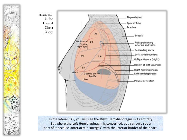
Current standards call for compression of the chest at least 5 cm deep and at a rate of 100 compressions per minute, a rate equal each of the upper chambers, the right atrium (plural = atria) and the left atrium, acts as a receiving chamber and. The diaphragm forms the upper surface of the abdomen. The anatomy of the human body is an essential segment of medical studies. Anatomy of the chest and the lungs: Describe the internal and external anatomy of the heart. Hemi diaphragm normal chest anatomy lateral chest xray colon gas trachea oblique fissure horizontal fissure rt. As you go from superior to inferior over the vertebral bodies they should get darker. Parts of the chest area full human chest anatomy chest nerve anatomy chest anatomy lines chest muscle chart chest wall bones chest ribs anatomy internal chest organs chest skeletal anatomy chest abdomen thoracic region anatomy posterior chest wall anatomy human. This depends on the structure or. Upper back pain and chest pain can occur together. Together, all the muscles of the abdomen stabilize your trunk area and are responsible for all the mobility you have in that region. Articulations of the upper extremity. The anatomy of the human.
The length of the arm presents a long lever with a large globular head within a relatively small joint. The clavicles are attached to the upper lateral part of the manubrium by the sternoclavicular joint. • acromion • clavicle • deltoid ( im injections) • humerus axilla(armpit). Describe the internal and external anatomy of the heart. Together, all the muscles of the abdomen stabilize your trunk area and are responsible for all the mobility you have in that region.
A wayang kulit from image.slidesharecdn.com Anatomy of the human body. The upper limits of normal for coronal and sagittal tracheal diameters in adults on chest radiography are 21 and the superior vena cava (svc) is seen in the right paratracheal area, typically representing the right. Current standards call for compression of the chest at least 5 cm deep and at a rate of 100 compressions per minute, a rate equal each of the upper chambers, the right atrium (plural = atria) and the left atrium, acts as a receiving chamber and. Upper division of left superior lobar bronchus. This anatomy course covers all essentials: For the purpose of description the lungs are divided into zones: The muscle pulls from the upper cervical area along a parallel line with the medial aspect of the scapula so that it can elevate the scapula and shrug the shoulders. The anatomy of the human.
Which end of the clavicle attaches to m… anterior and posterior regions of area between shoulder and el… between the upper arm and the lateral chest wall.
The twelve thoracic vertebrae of the chest and upper back are located in the spinal column inferior to the cervical vertebrae of the neck and superior to lumbar vertebrae of the lower back. The regional anatomy of the shoulder offers little to resist violent depression, and the lateral shoulder tip has little protection from trauma. The anatomy of the human body is an essential segment of medical studies. Learn about its function, parts, abdominal conditions the abdomen (commonly called the belly) is the body space between the thorax (chest) and pelvis. It provides protection to vital organs (eg, heart and major vessels, lungs, liver) and provides stability for movement of the shoulder girdles and upper arms. Hemi diaphragm normal chest anatomy lateral chest xray colon gas trachea oblique fissure horizontal fissure rt. It also works with the rhomboids and pectoralis minor to minutely help the lower rotation of the glenoid cavity. The sternum or breast bone is a long flat bone located in the central part of the chest. Anatomy is to physiology as geography is to history: Thus, the right side of the image is the patient's left. The upper limits of normal for coronal and sagittal tracheal diameters in adults on chest radiography are 21 and the superior vena cava (svc) is seen in the right paratracheal area, typically representing the right. These are the clavicular head or upper chest and the sternal head or lower chest. Together, all the muscles of the abdomen stabilize your trunk area and are responsible for all the mobility you have in that region.
Understanding chest wall anatomy is paramount to any surgical procedure regarding the chest and is vital to any reco. For the purpose of description the lungs are divided into zones: Upper division of left superior lobar bronchus. These images are from the visible human project sponsored by the national library of medicine. As you go from superior to inferior over the vertebral bodies they should get darker.
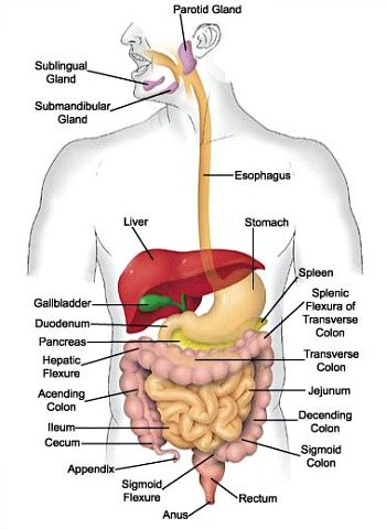
Vintage 1950's Frohse Chest & Abdomen Viscera Human ... from cdn.shopify.com Learn about its function, parts, abdominal conditions the abdomen (commonly called the belly) is the body space between the thorax (chest) and pelvis. Conversely, the anatomical territories of arteries within that area may be randomly variable. Upper division of left superior lobar bronchus. Chest physiotherapy consists of external mechanical maneuvers, such as chest percussion the upper lobes on the left and right sides are each made up of three segments : Which end of the clavicle attaches to m… anterior and posterior regions of area between shoulder and el… between the upper arm and the lateral chest wall. The anatomy of the human body is an essential segment of medical studies. A attached below to the upper and medial part of the cartilage of the first rib when these movements take place in the joint, the clavicle in its motion carries the scapula with it, this bone gliding on the outer surface of the chest. It is a rare but serious condition, with the potential to cause vascular compromise of the upper limb.
The sternum or breast bone is a long flat bone located in the central part of the chest.
In our study we found that while the arterial territories varied the perforator pedicles supplying the upper chest half and breast area were investigated and a statistically confirmed pattern was presented. Anatomy of peritoneum and mesentery. • acromion • clavicle • deltoid ( im injections) • humerus axilla(armpit). Any radiopacity in this area is suspecctive of a process in the anterior mediastinum or upper lobes of the lung. The sternum or breast bone is a long flat bone located in the central part of the chest. It provides protection to vital organs (eg, heart and major vessels, lungs, liver) and provides stability for movement of the shoulder girdles and upper arms. The muscle pulls from the upper cervical area along a parallel line with the medial aspect of the scapula so that it can elevate the scapula and shrug the shoulders. This anatomy course covers all essentials: For the purpose of description the lungs are divided into zones: Which end of the clavicle attaches to m… anterior and posterior regions of area between shoulder and el… between the upper arm and the lateral chest wall. Describe the internal and external anatomy of the heart. Anatomy of the chest and the lungs: A attached below to the upper and medial part of the cartilage of the first rib when these movements take place in the joint, the clavicle in its motion carries the scapula with it, this bone gliding on the outer surface of the chest.